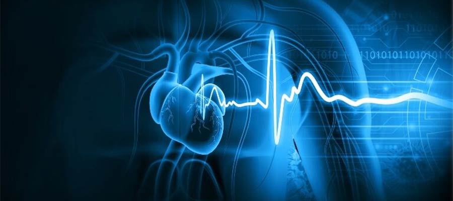
Electrocardiogram (ECG or EKG) Testing in Miami

Are you looking for Electrocardiogram (ECG or EKG) testing in Miami? Wellcare Med Group can perform Electrocardiogram (ECG or EKG) testing and other diagnostic tests related to neurological health whether your healthcare provider suggested it or an initiative you have taken on your own.
An electrocardiogram, called an ECG or EKG, is a medical test doctors use to check how your heart is working. This test measures the electrical signals that your heart produces as it beats. In this article, we’ll explain an ECG, how it works, the test, and why it’s essential for keeping your heart healthy.
What is an Electrocardiogram (ECG or EKG)?
An electrocardiogram (ECG or EKG) is a test that shows the electrical activity of your heart. It helps doctors see if your heart is beating normally and if it’s healthy. The terms ECG and EKG are different ways of saying the same thing—EKG is used in German, and ECG is used in English.
How Does an ECG Work?
The Heart’s Electrical System
Your heart beats because of electrical signals. These signals tell the heart muscles when to squeeze (contract) and when to relax. Here’s a simple way to understand how this works:
- The Sinoatrial (SA) Node is like your heart’s natural battery. It sends out electrical signals that start each heartbeat.
- Atrioventricular (AV) Node: This part of the heart passes the signal from the top part of your heart to the bottom, helping it beat in a coordinated way.
- His-Purkinje Network: These tiny wires spread the electrical signal through your heart, causing it to beat and pump blood.
Recording Electrical Activity
An ECG records the electrical signals your heart produces. Small, sticky patches called electrodes are placed on your skin, picking up the signals and sending them to a machine. The machine then draws a picture of these signals on paper or a screen, showing how your heart works.
The ECG Procedure
Getting Ready for the Test
Before an ECG, you may need to remove jewelry or any clothing above your waist to place the electrodes on your skin. The doctor or nurse will clean the spots where the electrodes go to ensure they stick well.
Where the Electrodes Are Placed
Electrodes are small sticky patches on your chest, arms, and legs. These patches pick up the electrical signals from your heart.
Taking the Test
Once the electrodes are in place, they are connected to the ECG machine with wires. The machine then records your heart’s electrical activity, and you’ll see it as lines and waves on a screen or paper.
How Long Does It Take?
The whole process usually takes about 5 to 10 minutes. The recording only takes a few seconds, but setting everything up takes longer.
Understanding the ECG Results
The ECG Graph
The ECG machine shows your heart’s activity as a series of waves:
- P Wave: This wave shows when the top parts of your heart (atria) contract.
- QRS Complex: This is the biggest wave and shows when the bottom parts of your heart (ventricles) contract.
- T Wave: This shows when the ventricles relax.
- U Wave (sometimes there): This is a small wave that sometimes appears, and it’s related to the relaxation of other heart muscles.
Important Parts of the ECG
The ECG graph also has important sections called intervals and segments:
- PR Interval: The time it takes for the signal to go from the atria to the ventricles.
- QT Interval: The time it takes for the ventricles to contract and then relax.
- ST Segment: The flat part between the end of the contraction and the start of the relaxation.
Normal vs. Abnormal ECG
A normal ECG has a steady rhythm and consistent wave shapes. If there are any problems, like an irregular heartbeat or signs of a heart attack, the waves will look different, and a doctor can use these differences to figure out what might be wrong.
Why Do We Use ECGs?
Finding Heart Problems
An ECG is one of the first tests doctors use to determine if something is wrong with your heart. It can help detect:
- Coronary Artery Disease happens when blood doesn’t flow well to the heart muscle.
- Heart Attack: This is when part of the heart muscle is damaged due to a lack of blood flow.
- Cardiomyopathy: This is when the heart muscle becomes weak or stiff.
- Pericarditis: This is inflammation of the sac around the heart.
- Electrolyte Imbalances: These are problems with the minerals in your blood that can affect your heart.
Checking If Treatments Are Working
Doctors also use ECGs to see if treatments for heart problems are working. For example, if someone is taking medicine for their heart, an ECG can show if the medicine is helping.
Before Surgery
Sometimes, doctors will do an ECG before surgery to make sure your heart is healthy enough for the operation.
Limits of the ECG
Even though an ECG is a great tool, it has some limits:
- Short-Term Picture: An ECG only shows what’s happening with your heart. If you have a problem that comes and goes, it might not show up on the test.
- Heart Structure: An ECG doesn’t show the actual structure of your heart. If doctors need to see the heart, they might do an ultrasound or another imaging test.
- Expert Interpretation: The ECG results can be tricky to read, so a trained doctor must look at them to make sure the diagnosis is correct.
Conclusion
An electrocardiogram (ECG or EKG) is a simple, quick, and important test that helps doctors understand how your heart works. Recording your heart’s electrical signals can show whether your heart is beating normally or if there might be a problem. While it’s imperfect and can’t show everything about your heart, the ECG is critical in keeping your heart healthy and diagnosing any issues early.
Whether getting an ECG as part of a routine check-up, diagnosing a heart problem, or monitoring treatment, understanding how the test works and what it can tell you about your heart is vital for your health.
